Study Reveals Subtle Neuroimaging Differences After Late Preterm Birth
A new longitudinal investigation has identified subtle neuroimaging differences in children born late preterm compared to those born full-term. Researchers utilized data from the U.S.-based Adolescent Brain Cognitive Development Study to examine the long-term neurodevelopmental characteristics of children aged nine to 11 years old. The findings, published in Frontiers in Pediatrics, were led by a team from the University of Fukui in Japan, including specially appointed Professor Akemi Tomoda.
Evaluating Late Preterm Birth Outcomes
Late preterm birth is defined as delivery occurring between 34 and 36 weeks of gestation. This specific window coincides with a period of rapid brain development characterized by substantial white matter expansion, synapse formation, dendritic growth, and cortical folding. Although previous investigations have reported mixed results regarding the long-term implications of being born several weeks early, the clinical picture has remained largely unclear.
To explore this further, the research team compared 1,093 children born late preterm with 8,984 children born full-term. The analysis evaluated behavioral and emotional problems, cognitive function, structural magnetic resonance imaging, diffusion tensor imaging, and resting-state functional magnetic resonance imaging. Overall, the longitudinal analysis found no broad differences between the groups regarding behavioral and emotional problems, most cognitive outcomes, cortical thickness, cortical surface area, or resting-state functional connectivity.
Structural MRI and Diffusion Tensor Imaging Findings
Structural MRI identified group differences in six regional brain volumes, including the left postcentral, right fusiform, right middle temporal, right pars orbitalis, right supramarginal, and right insular regions. However, the study noted that these particular measures did not show significant timepoint or group-by-timepoint effects after false discovery rate correction was applied.
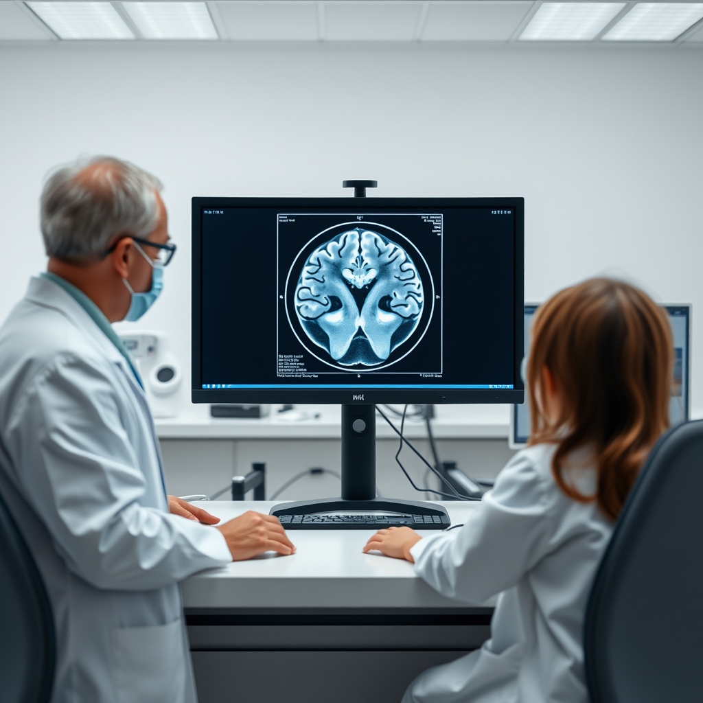
The clearest differences emerged from diffusion tensor imaging, a technique that assesses the diffusion properties of brain tissue. Group effects were identified in selected white matter, cortical, and subcortical fractional anisotropy and mean diffusivity measures. Furthermore, group-by-timepoint interactions were detected across several white matter tracts and selected cortical and subcortical regions. According to the analysis, many of these interactions reflected decreases in diffusion tensor imaging measures among full-term children over a two-year period, whereas changes among late preterm children were smaller, nonsignificant, or less consistent.
- No widespread differences in behavioral or emotional problems
- Regional variations found in selected brain volumes and diffusion measures
- Longitudinal changes showed distinct trajectories over two years
Study Limitations and Clinical Interpretation
Lead researcher Akemi Tomoda noted that the findings suggest the effects of late preterm birth may be subtle and do not necessarily manifest as broad behavioral or cognitive difficulties. Instead, brain imaging may reveal regional differences in maturation that are not immediately evident in everyday functioning. The absence of widespread behavioral or cognitive differences is considered reassuring for families.
Importantly, the authors emphasized several key limitations. The observational design, retrospective gestational-age reporting, substantial follow-up attrition, and the presence of only two assessment timepoints limit the interpretation of the results. Additionally, because fractional anisotropy and mean diffusivity are influenced by multiple tissue properties, the findings do not establish a specific biological mechanism, such as altered myelination or axonal organization. Furthermore, the imaging differences do not establish that every child born late preterm will develop difficulties or that these variations directly cause functional problems. Further longitudinal research is required to determine whether these subtle neuroimaging differences persist, change, or relate to later educational and clinical outcomes.