Human Brain Actually Consists of Two Separate Organs, Landmark Study Reveals
For centuries, the human brain has been conceptualized as a single, unified organ. However, groundbreaking research led by Stanford Medicine reveals that what we routinely call the brain is actually two distinct nervous systems packaged together. Published in the journal Nature Neuroscience, the findings overturn a longstanding model of human development and could pave the way for new treatments targeting debilitating brain stem conditions.
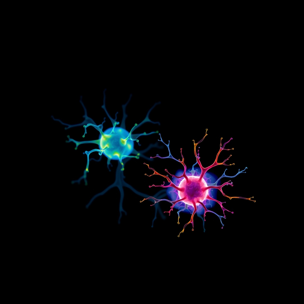
For decades, neuroscientists subscribed to the theory that a single progenitor cell early in embryonic development gave rise to the entire brain, meaning all parts shared a common developmental origin. The new study demonstrates that the human brain instead consists of two ancient systems that evolved independently over hundreds of millions of years: a primitive region that regulates automatic functions like breathing and heartbeat, and a more recent structure responsible for higher-level cognition, mathematics, and abstract reasoning.
Kyle Loh, Ph.D., associate professor of developmental biology at Stanford and senior author of the study, explained that the front of the brain arises from an entirely different progenitor cell than the back of the brain. Graduate students Carolyn Dundes and Rayyan Jokhai served as co-first authors on the research project.
Understanding the Split: Forebrain Versus Hindbrain
To understand the significance of the discovery, researchers point to the distinct jobs handled by the adult brain’s primary regions:
- The forebrain and midbrain: Manage higher-level thinking, language, consciousness, and abstract reasoning.
- The hindbrain: Located at the back of the skull and frequently called the brain stem, it controls essential, life-sustaining functions such as sleeping, breathing, heartbeat regulation, hunger urges, and the muscles of the face, throat, and tongue involved in swallowing and speech.
Despite the crucial role of the hindbrain, scientists have struggled for decades to generate human hindbrain neurons in a laboratory setting. This limitation severely hampered investigation into severe medical conditions originating in the brain stem, such as amyotrophic lateral sclerosis (ALS, also known as Lou Gehrig’s disease) and spinal muscular atrophy (SMA), which is a leading genetic cause of death in infants under one year old. In both disorders, hindbrain neurons gradually fail, robbing patients of the ability to swallow and breathe.
According to the Stanford team, previous attempts to culture these cells likely failed because researchers attempted to coax forebrain and hindbrain progenitors into becoming cells they were fundamentally incapable of forming.
Tracing Embryonic Origins and DNA Packaging
The research breakthrough originated from examining the earliest moments of embryonic development during gastrulation—the stage when the body first establishes its shape. Investigating developing mouse embryos, the Stanford investigators discovered that the hindbrain follows a separate developmental path running parallel to, rather than branching off from, the pathway creating the forebrain and midbrain.
The team identified two distinct, mutually exclusive brain progenitor cell populations:
- Cells expressing the Otx2 gene, which are destined to form the forebrain and midbrain.
- Cells expressing the Gbx2 gene, which are committed exclusively to forming the hindbrain.
Furthermore, the researchers analyzed the chromatin—the DNA packaging that determines which genes remain accessible—in these populations. They found that the anterior neural ectoderm (future forebrain and midbrain) and posterior neural ectoderm (future hindbrain) feature fundamentally different chromatin configurations. These distinct configurations lock each progenitor cell into its specific developmental fate, much like two trains traveling on parallel tracks that never intersect.
Growing Hindbrain Neurons in the Lab
Armed with this developmental insight, the researchers successfully coaxed human pluripotent stem cells—versatile cells capable of generating any cell type in the human body—into becoming functional hindbrain motor neurons in a petri dish for the very first time. These laboratory-grown neurons exhibited authentic hallmarks of hindbrain cells, including waves of electrical activity known as action potentials and the production of specific proteins associated with facial and swallowing muscle control.
Expanding their perspective across evolutionary history, the investigators identified the same two-origin brain pattern in chickens, zebrafish, and even acorn worms—small ocean-floor creatures sharing a distant common ancestor with humans. This suggests that evolution physically pushed two existing, ancient neural systems together spatially.
The ability to grow these specialized neurons in a laboratory environment provides researchers with a vital model to investigate what goes wrong in conditions like ALS and SMA, where obtaining living brain stem tissue was previously nearly impossible. Additionally, researchers note potential connections to metabolic studies, as the hindbrain houses the neural circuits responsible for regulating hunger—the exact biological pathway targeted by modern weight-loss medications such as semaglutide.
Source: Medical Xpress